Pneumonia Physiotherapy Treatment | Arunalaya Healthcare post thumbnail image

Best Pneumonia Physiotherapy Treatment in Delhi

What is Pneumonia Physiotherapy?

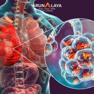
Pneumonia is an inflammatory condition of the lung parenchyma (the air sacs, or alveoli, and the surrounding tissue) primarily affecting the gas exchange units. It is characterized by inflammation and fluid or pus filling the alveoli, which hinders the normal exchange of oxygen and carbon dioxide.

Causes of Pneumonia

Pneumonia can be caused by various infectious agents, and less commonly by non-infectious factors. The most common causes include:-

  • Bacteria (Most Common Cause in Adults):
    • Streptococcus pneumoniae (Pneumococcus): The most common bacterial cause.
    • Haemophilus influenzae: Another common cause, especially in individuals with underlying lung disease.
    • Staphylococcus aureus: Can cause severe pneumonia, often after a viral illness or in hospitalized patients (e.g., MRSA).
    • Mycoplasma pneumoniae: Causes “walking pneumonia,” a milder form.
    • Chlamydia pneumoniae: Another cause of atypical pneumonia.
    • Klebsiella pneumoniae: More common in individuals with chronic lung disease or alcoholism.
  • Viruses (Most Common Cause in Children and Infants):
    • Influenza virus (Flu): A very common cause, sometimes leading to secondary bacterial pneumonia.
    • Respiratory Syncytial Virus (RSV): A common cause in young children.
  • Adenovirus, Parainfluenza virus.
    • COVID-19 (SARS-CoV-2): A significant cause of viral pneumonia with potentially severe complications.
  • Fungi (Less Common, More in Immunocompromised Individuals):
    • Pneumocystis jirovecii (PJP): Common in people with HIV/AIDS.
    • Histoplasma capsulatum, Coccidioides immitis, Blastomyces dermatitidis: Found in specific geographic regions.
  • Other Causes (Non-Infectious):
    • Aspiration Pneumonia: Occurs when food, drink, vomit, or saliva is inhaled into the lungs instead of being swallowed. Common in individuals with swallowing difficulties (dysphagia), neurological conditions (stroke, Parkinson’s), or impaired consciousness.
    • Chemical Pneumonia: Inhalation of toxic gases, chemicals, or foreign materials.
    • Drug-Induced Pneumonia: Certain medications can cause lung inflammation.
    • Radiation Pneumonia: After radiation therapy to the chest for cancer.
Risk Factors for Pneumonia
  • Age (very young children and older adults >65)
  • Weakened immune system (HIV/AIDS, organ transplant, chemotherapy, chronic steroid use)
  • Chronic medical conditions (diabetes, heart disease, kidney disease, liver disease)
  • Chronic lung diseases (COPD, asthma, cystic fibrosis)
  • Smoking and excessive alcohol consumption
  • Dysphagia or neurological disorders affecting swallowing
  • Hospitalization, especially in ICUs or on ventilators (ventilator-associated pneumonia – VAP)
  • Malnutrition
Symptoms of Pneumonia

Symptoms can range from mild to severe, depending on the cause, age, overall health, and extent of lung involvement.

  • Cough: Often productive (producing phlegm or mucus) which may be green, yellow, or even bloody. Can also be dry.
  • Fever: High fever is common, sometimes with chills and shivering.
  • Shortness of Breath (Dyspnea): Difficulty breathing, especially with exertion.
  • Chest Pain: Sharp or stabbing pain that worsens with deep breaths or coughing (pleuritic pain).
  • Fatigue and Weakness: General feeling of malaise and exhaustion.
  • Sweating
  • Loss of Appetite
  • Nausea and Vomiting or Diarrhea: Less common, but can occur, especially with viral or atypical pneumonias.
  • Headache
  • Confusion or Delirium: More common in older adults or those with severe infections.
  • Bluish discoloration of lips or fingernails (Cyanosis): A sign of severe oxygen deprivation.
Advanced Physiotherapy for Pneumonia

Physiotherapy plays a vital role in the management of pneumonia, particularly in hospital settings and for patients with severe symptoms or underlying respiratory conditions.

  • Respiratory Assessment: Auscultation (listening to breath sounds for crackles, diminished sounds), percussion, palpation, and observation of breathing patterns (rate, depth, accessory muscle use).
    • Oxygen Saturation (SpO2): Monitoring oxygen levels.
    • Cough Assessment: Efficacy of cough, sputum production (amount, color, consistency).
    • Pain Assessment: Chest pain, musculoskeletal pain.
    • Functional Capacity: Assessment of mobility, strength, and endurance.
    • Medical History: Reviewing underlying conditions, medications, and imaging (X-rays, CT scans).
  • Airway Clearance Techniques (ACTs): These are central to physiotherapy for pneumonia.
    • Manual Techniques
    • Percussion/Clapping: Rhythmic striking of the chest wall over the affected lung segment to dislodge mucus.
    • Vibrations/Shaking: Gentle, sustained pressure applied to the chest wall during exhalation to move secretions towards larger airways.
    • Postural Drainage: Positioning the patient to allow gravity to drain secretions from specific lung segments into the main airways, where they can be coughed up. This requires knowledge of lung anatomy and segment positioning.
    • Active Cycle of Breathing Techniques (ACBT): A patient-led technique involving:
    • Breathing Control: Gentle breathing to relax airways.
    • Thoracic Expansion Exercises: Deep breaths to get air behind secretions.
    • Forced Expiration Technique (FET)/”Huffing”: Short, sharp expirations (like steaming up a mirror) to move secretions from peripheral to central airways, followed by a cough.
    • Autogenic Drainage: A self-drainage technique using varying depths and speeds of breathing to mobilize secretions from peripheral to central airways without forced coughing.
  • Positive Expiratory Pressure (PEP) Devices:
    • PEP Mask/Valve: Breathing out through a device that provides resistance, creating positive pressure in the airways, which helps to stent airways open and move secretions.
    • Oscillatory PEP (e.g., Acapella, Flutter valve): Combines PEP with oscillations to vibrate secretions and make them easier to clear.
    • Assisted Cough Techniques: For patients with weak coughs, manual assistance (e.g., abdominal thrust) to make the cough more effective.
  • Breathing Exercises and Re-education
    • Diaphragmatic Breathing (Abdominal Breathing): Teaching patients to use their diaphragm more effectively to reduce the work of breathing and improve ventilation, especially in the lower lung lobes.
    • Pursed-Lip Breathing: Breathing out slowly through pursed lips helps to keep airways open, reduce air trapping, and control shortness of breath.
    • Segmental Breathing: Directing breath to specific areas of the lungs to improve ventilation in under-inflated segments.
    • Breathing Control Strategies: Teaching patients how to manage breathlessness during activity.
  • Mobility and Exercise Progression-
    • Early Mobilization: As soon as medically stable, encouraging sitting up, standing, and walking, even short distances. This helps to improve lung expansion, prevent deconditioning, and reduce the risk of secondary complications like deep vein thrombosis (DVT).
    • Graded Exercise Program: Progressing from bed exercises to ambulation, then increasing intensity and duration of activity as tolerance improves. This combats deconditioning and improves overall functional capacity.
    • Incentive Spirometry: Using a device to encourage deep breaths and lung expansion, preventing atelectasis (lung collapse).
  • Postural Management
    • Optimizing body positioning to promote lung expansion and comfort. This includes frequent position changes to prevent pressure sores and improve ventilation distribution.
    • Head of bed elevation to reduce aspiration risk.


BOOK AN APPOINTMENT

Working Hours

Mon - Sat: 9:00AM to 8:30PM
Sunday: 9:30AM to 7:30PM

Call Us

+91 8090080906
+91 8090080907
+91 8866991000


    Add Your Heading Text Here