Posterior Tibial Tendon Dysfunction | Arunalaya Healthcare post thumbnail image

Best Posterior Tibial Tendon Dysfunction Treatment in Delhi

What is Posterior Tibial Tendon Dysfunction ?

Posterior Tibial Tendon Dysfunction (PTTD), also commonly known as Adult Acquired Flatfoot Deformity (AAFD), is a progressive and often painful condition that results from changes in the posterior tibial tendon. This tendon is crucial for supporting the arch of the foot and helping it function during walking. When it becomes impaired, the arch can gradually flatten, leading to significant mobility limitations and pain.

Causes of Posterior Tibial Tendon Dysfunction

PTTD is primarily an overuse injury, where the tendon gradually weakens and degenerates over time, rather than a sudden acute injury. Several factors contribute to its development:-

  • Repetitive Microtrauma: The most common cause is chronic repetitive loading and microtrauma to the tendon, often from activities like running, walking, hiking, or climbing stairs, especially in individuals with pre-existing foot abnormalities.
  • Anatomical Factors: The posterior tibial tendon makes an acute turn around the medial malleolus (inner ankle bone), placing significant tension on the tendon in this region, which can contribute to its degeneration. A hypovascular (poor blood supply) region distal and posterior to the medial malleolus may also play a role.
  • Degenerative Changes: The condition is often characterized by tendinosis (degeneration of the tendon tissue) rather than pure inflammation (tendinitis), though inflammation can be present, especially in earlier stages.
  • Risk Factors:
    • Age: Most common in individuals over 40.
    • Gender: More prevalent in females.
    • Obesity: Increased weight places additional strain on the foot and ankle structures.
    • Diabetes and Hypertension: These systemic conditions can affect tendon health, leading to decreased elasticity and increased risk of degeneration.
    • Previous Trauma: Acute injuries, such as ankle sprains or fractures, can predispose individuals to PTTD.
    • Pre-existing Flatfoot/Over-pronation: Feet that are already somewhat flat or tend to roll inward excessively (over-pronation) place greater chronic stress on the posterior tibial tendon.
    • Poor Footwear: Lack of adequate arch support or wearing unstable shoes (e.g., flip-flops) can exacerbate the condition.
    • High-Impact Sports/Activities: Engaging in activities that involve repetitive heavy impact on the feet.
Symptoms of Posterior Tibial Tendon Dysfunction

Symptoms of PTTD typically develop gradually and progress as the condition worsens. The presentation is often categorized into stages:-

  • Stage I (Inflammation/Early Dysfunction):
    • Pain and tenderness along the inside of the foot and ankle, often behind or below the medial malleolus (inner ankle bone).
    • Swelling in the same area.
    • Pain often worsens with activity, prolonged walking, standing, or at the end of the day.
    • Arch may still appear normal or only slightly flattened.
    • Patient can still perform a single-leg heel raise (standing on tiptoes on the affected foot), though it may be painful.
    • Mild weakness with inversion of the foot (turning the sole inward).
  • Stage II (Progressive Deformity)
    • The arch begins to flatten noticeably (adult-acquired flatfoot).
    • The heel may start to shift outward (valgus deformity).
    • The front of the foot may appear abducted (turning outward), leading to a “too many toes” sign (when viewed from behind, more toes are visible on the affected foot compared to the unaffected foot).
    • Significant difficulty or inability to perform a single-leg heel raise.
    • Pain may still be on the inside of the foot, but as the deformity progresses, pain can shift to the outside of the ankle due to impingement of the fibula against the calcaneus.
    • Increased weakness of the posterior tibial muscle.
  • Stage III (Fixed Deformity and Arthritis):
    • The flatfoot deformity becomes rigid or fixed, meaning it cannot be corrected manually.
    • Significant arch collapse and severe valgus heel.
    • Arthritis may develop in the foot and ankle joints due to abnormal stresses.
    • Pain may be severe and widespread, affecting multiple areas of the foot and ankle.
    • Pronounced difficulty with walking and daily activities.
  • Stage IV (Ankle Arthritis):
    • Deformity extends to the ankle joint, leading to ankle arthritis.
    • Severe pain and functional limitations.
Advanced Physiotherapy for Posterior Tibial Tendon Dysfunction

Assessment:

    • Gait Analysis: Observing walking pattern, pronation, and compensatory movements.
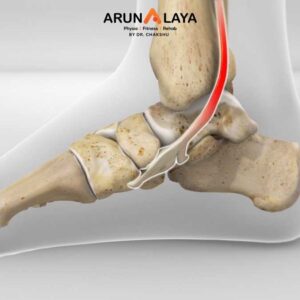
    • Strength Testing: Assessing the strength of the posterior tibial muscle (inversion and plantarflexion) and other surrounding muscles (peroneals, calf, hip muscles).
    • Range of Motion (ROM) Assessment: Measuring ankle, subtalar, and midfoot joint mobility.
    • Palpation: Identifying tender areas along the tendon and surrounding structures.
    • Special Tests: “Too many toes” sign, single-leg heel raise test (assessing ability and quality of movement).
    • Balance and Proprioception Assessment.

Advanced Physiotherapy

  • Pain and Inflammation Management (Acute/Early Stages):
    • Relative Rest/Activity Modification: Reducing activities that aggravate the tendon.
    • Cryotherapy (Ice): To reduce pain and swelling.
    • Modalities: Therapeutic ultrasound or other electrophysical agents may be used to aid tissue healing and reduce pain.
    • Manual Therapy for Edema Control: Gentle lymphatic drainage techniques if significant swelling is present.

Advanced Therapeutic Exercise:-

  • Posterior Tibial Tendon Strengthening (Graded Progressive Overload):
    • Isometrics: Initial focus on gentle contractions without movement, especially in painful stages.
    • Concentric Exercises: Strengthening the muscle as it shortens (e.g., controlled ankle inversions with resistance bands).
    • Eccentric Exercises: Crucial for tendon health. Strengthening the muscle as it lengthens (e.g., slowly lowering the heel from a heel raise position, emphasizing control during the “descent”). This is vital for improving the tendon’s capacity to tolerate load.
    • Heel Raises: Progressing from double-leg to single-leg heel raises, focusing on controlled movement and proper alignment (avoiding foot collapse). May include elevated heel raises or weighted heel raises as strength improves.
    • Active Arch Lifts: Exercises to strengthen the intrinsic foot muscles and encourage active arch support (e.g., “short foot” exercise, towel scrunches).
    • Calf Stretching: Addressing tightness in the gastrocnemius and soleus muscles (calf muscles), which can increase stress on the posterior tibial tendon.
    • Proximal Strengthening: Strengthening hip abductors, extensors, and external rotators to improve lower limb alignment and reduce compensatory movements at the foot and ankle (e.g., glute bridges, clam shells, side leg raises).

Balance and Proprioception Training:

  • Progressing from stable to unstable surfaces (e.g., foam pad, wobble board).
    • Single-leg standing, dynamic balance exercises, and functional reaching drills.
    • Gait Retraining: Analyzing and correcting abnormal walking patterns to reduce stress on the affected tendon and promote more efficient movement.
    • Plyometrics/Agility (Advanced Stages, if appropriate): For active individuals, carefully graded jumping, hopping, and agility drills may be introduced to prepare for higher-impact activities, but only after significant strength and stability have been restored.
  • Manual Therapy Techniques (Hands-On Approach):

Manual therapy plays a significant role in PTTD by addressing soft tissue restrictions, improving joint mobility, and managing pain.

Soft Tissue Mobilization:

 Deep Transverse Friction Massage: Applied directly to the posterior tibial tendon (if not acutely inflamed) to stimulate healing, break down adhesions, and improve tissue extensibility.

Myofascial Release: Techniques applied to the calf muscles, plantar fascia, and other surrounding fascial tissues to release tension and improve overall lower limb mechanics.

  •  Instrument-Assisted Soft Tissue Mobilization (IASTM): Using specialized tools (e.g., Graston, HawkGrips) to detect and treat fascial restrictions and scar tissue more effectively.

 Joint Mobilization:

  • Subtalar Joint Mobilization: Gentle accessory glides and oscillations to improve mobility in the subtalar joint, which is often restricted in PTTD due to collapse and muscle guarding.
  • Midfoot Joint Mobilization: Mobilizing joints like the talonavicular, calcaneocuboid, and cuneiform joints to restore proper foot mechanics and reduce stiffness.
  • Ankle Joint Mobilization: To ensure optimal dorsiflexion and plantarflexion range.
  • Manual Stretching: Therapist-assisted stretches for tight calf muscles, Achilles tendon, and other restricted areas.
  • Trigger Point Release: Addressing painful muscle knots in the calf or foot muscles that may be contributing to pain or altered mechanics.

BOOK AN APPOINTMENT

Working Hours

Mon - Sat: 9:00AM to 8:30PM
Sunday: 9:30AM to 7:30PM

Call Us

+91 8090080906
+91 8090080907
+91 8866991000


    Add Your Heading Text Here