Tarsal Tunnel Syndrome | Arunalaya Healthcare post thumbnail image

Best Tarsal Tunnel Syndrome Treatment in Delhi

What is Tarsal Tunnel Syndrome ?

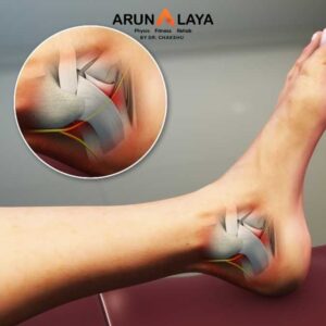
Tarsal Tunnel Syndrome (TTS) is a condition characterized by compression or squeezing of the posterior tibial nerve as it passes through a narrow anatomical space in the ankle called the tarsal tunnel. This condition is often referred to as the ankle’s counterpart to carpal tunnel syndrome in the wrist.

  • The Tarsal Tunnel-The tarsal tunnel is a confined space located on the inside of the ankle, just below the medial malleolus (the bony prominence on the inner side of the ankle). It is formed by:-
    • Bones: The medial malleolus, talus, and calcaneus.
    • Ligaments: Primarily the flexor retinaculum, a strong fibrous band that arches over these bones, forming the “roof” of the tunnel.
  • Through this tunnel pass several vital structures, including:-
    • The posterior tibial nerve (the main nerve involved in TTS).
    • The posterior tibial artery and veins.
    • Tendons of several muscles: Tibialis posterior, Flexor digitorum longus, and Flexor hallucis longus.
Causes of Tarsal Tunnel Syndrome

Any condition that reduces the space within the tarsal tunnel or directly irritates/compresses the posterior tibial nerve can lead to TTS

  •  Space-Occupying Lesions (Intrinsic Causes):
    • Ganglion cysts: Fluid-filled sacs.
    • Lipomas: Fatty tumors.
    • Varicose veins: Enlarged veins.
    • Tenosynovitis: Inflammation of the tendon sheaths (e.g., of the tibialis posterior tendon).
    • Osteophytes/Bone spurs: Bony outgrowths, often due to arthritis.
    • Perineural fibrosis: Thickening or scarring around the nerve.
    • Accessory muscles: An extra muscle belly within the tunnel.
  • Biomechanical Factors (Extrinsic Causes):
    • Flat feet (Pes Planus) or Pronated Feet: This is a very common contributing factor. When the arch collapses and the foot pronates excessively, it can stretch and compress the tibial nerve as it passes through the tunnel. The heel may also tilt outward, putting tension on the nerve.
    • High arches (Pes Cavus): Less common, but can also alter foot mechanics and nerve tension.
    • Repetitive stress or overuse: Activities that involve repetitive ankle movements, prolonged standing, or running can irritate the nerve over time.
  • Trauma:
    • Ankle sprains (especially inversion sprains): Swelling, scar tissue formation, or even direct nerve contusion from an ankle injury can trigger TTS.
    • Fractures or dislocations of the ankle or foot bones.
  • Systemic Diseases:
    • Diabetes: Can cause peripheral neuropathy, making nerves more susceptible to compression and damage.
    • Arthritis: Conditions like rheumatoid arthritis or osteoarthritis can cause inflammation and swelling within the joint and surrounding tissues, compressing the nerve.
    • Hypothyroidism.
    • Gout.
    • Idiopathic: In some cases, no clear cause can be identified.
Symptoms of Tarsal Tunnel Syndrome

Symptoms typically affect the inside of the ankle and the bottom of the foot, radiating towards the toes. They often worsen with activity, prolonged standing, or at night.

  • Pain: Can be sharp, shooting, aching, or burning. It is usually felt on the inside of the ankle and the sole of the foot, but can radiate into the arch, toes, and sometimes even up into the calf.
  • Numbness: A loss of sensation in parts of the sole of the foot and toes.
  • Tingling or “pins and needles” (paresthesia): A prickly or buzzing sensation.
  • Burning sensation: Especially on the bottom of the foot.
  • Weakness: In the small muscles of the foot (intrinsic foot muscles) that the tibial nerve supplies, leading to difficulty with toe movements or foot stability.
  • Symptoms worsen with activity: Pain and other symptoms often increase with walking, running, or standing for long periods. They may be relieved by rest.
  • Tinel’s Sign: A classic diagnostic test where tapping over the tarsal tunnel (behind the medial malleolus) reproduces or exacerbates the tingling, burning, or pain in the distribution of the nerve.
  • Positive Dorsiflexion-Eversion Test: Extreme dorsiflexion and eversion of the foot for 5-10 seconds may reproduce symptoms by stretching the nerve.
Advanced Physiotherapy for Tarsal Tunnel Syndrome
  • Reduce inflammation and nerve irritation.
  • Improve mobility of the nerve (nerve gliding).
  • Correct biomechanical imbalances (e.g., flat feet).
  • Strengthen supportive musculature.
  • Patient education and activity modification.

Advanced Physiotherapy

  • Manual Therapy:-
    • Nerve Gliding (Neural Mobilization): This is a cornerstone of TTS physiotherapy. The therapist uses specific, gentle techniques to “mobilize” the posterior tibial nerve, encouraging it to glide more freely within the tarsal tunnel and along its path. This can involve movements of the ankle and foot that gently stretch and release tension on the nerve, such as ankle dorsiflexion combined with toe extension and eversion, performed rhythmically and pain-free.
  • Soft Tissue Mobilization:
    • Deep Tissue Massage/Myofascial Release: To the calf muscles (gastrocnemius, soleus), tibialis posterior, and intrinsic foot muscles to release tightness, reduce muscle spasms, and improve tissue mobility that might be compressing the nerve or contributing to biomechanical faults.
    • Scar Tissue Mobilization: If the TTS is post-traumatic (e.g., after an ankle sprain/fracture), specific techniques to break down adhesions around the nerve and within the tunnel.
    • Joint Mobilizations: While not directly manipulating the nerve, specific gentle mobilizations to the subtalar joint and midfoot joints can be crucial. If these joints are stiff, they can alter foot mechanics and increase tension on the posterior tibial nerve. Mobilizations aim to restore normal joint play, particularly for inversion/eversion, and improve overall foot flexibility. The ankle (talocrural) joint may also be mobilized to ensure optimal dorsiflexion.
    • Taping: Specific taping techniques (e.g., low-dye taping or arch support taping) can be used to temporarily support the arch and reduce pronation, providing immediate relief and allowing the patient to experience the benefits before a permanent orthotic.
  • Therapeutic Exercises:-
    • Stretching:
    • Calf Stretches: (Gastrocnemius and Soleus) to improve ankle dorsiflexion and reduce tension on the posterior tibial nerve pathway.
    • Plantar Fascia Stretches: To improve flexibility of the foot’s arch.
    • Achilles Tendon Stretches: To address any tightness that influences foot mechanics.
  • Strengthening:
    • Foot Intrinsic Muscles: Exercises like towel curls, marble pickups, and “short foot” exercises to strengthen the small muscles that support the arch and stabilize the foot.
    • Extrinsic Foot and Ankle Muscles: Strengthening exercises for the tibialis posterior (to support the arch), tibialis anterior (for dorsiflexion), and peroneal muscles (for eversion) using resistance bands or body weight.
    • Hip and Core Strengthening: To improve proximal stability and address any kinetic chain imbalances that might contribute to foot dysfunction.
  • Balance and Proprioception Training:
    • Progressive balance exercises on stable surfaces (single leg stance), then unstable surfaces (foam pads, wobble boards, BOSU balls) to improve ankle stability and neuromuscular control.
    • Dynamic balance activities (e.g., walking on uneven surfaces, tandem walking, agility drills) as pain allows.
  • Modalities :
    • Ice/Cold Therapy: For pain and inflammation, especially after activity.
    • TENS (Transcutaneous Electrical Nerve Stimulation): For pain modulation.
    • Ultrasound: For deep tissue healing and pain relief, though its effectiveness for nerve compression is debated.
    • Low-Level Laser Therapy (LLLT): May promote nerve healing and reduce inflammation.


BOOK AN APPOINTMENT

Working Hours

Mon - Sat: 9:00AM to 8:30PM
Sunday: 9:30AM to 7:30PM

Call Us

+91 8090080906
+91 8090080907
+91 8866991000


    Add Your Heading Text Here