Peroneal Tendon Subluxation: Causes & Rehab Guide post thumbnail image

Best Peroneal Tendon Subluxation Treatment in Delhi

What is Peroneal Tendon Subluxation ?

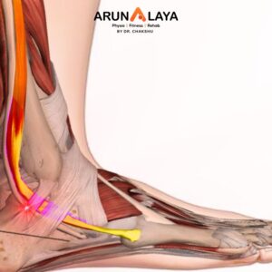
Peroneal tendon subluxation, also known as peroneal tendon dislocation or snapping ankle syndrome, occurs when one or both of the peroneal tendons (peroneus longus and peroneus brevis) slip out of their normal groove behind the lateral malleolus (the outer ankle bone). This is distinct from tendinopathy, which involves degeneration within the tendon itself, although subluxation can lead to tendinopathy over time due to chronic irritation.

Causes of Peroneal Tendon Subluxation

The primary cause is a disruption or laxity of the superior peroneal retinaculum (SPR), a fibrous band that holds the peroneal tendons securely in the groove behind the lateral malleolus.

Causes

  • Acute Trauma:
    • Forceful Ankle Dorsiflexion with Eversion/Inversion: This is the most common mechanism. A sudden, strong contraction of the peroneal muscles, often during activities like skiing, ice skating, or sports with rapid changes in direction (basketball, soccer, rugby), can cause the tendons to pull forcefully against the retinaculum, leading to a tear or avulsion (separation) of the SPR from the fibula.
    • Severe Ankle Sprain (especially inversion sprains): While typically associated with lateral ankle ligament injuries, a severe inversion sprain can sometimes injure the SPR, allowing the tendons to dislocate.
    • Direct Trauma: A direct blow to the outside of the ankle.
  • Anatomical Predisposition
    • Shallow Fibular Groove: The groove behind the lateral malleolus where the tendons sit might naturally be too shallow or even convex instead of concave, making it easier for the tendons to slip out.
    • Absent or Insufficient Fibular Ridge: The bony ridge that helps deepen the groove may be small or absent.
    • Congenital Laxity of the SPR: Some individuals may be born with a naturally looser superior peroneal retinaculum.
    • Low-Lying Peroneus Brevis Muscle Belly: If the muscle belly extends too far distally, it can occupy more space in the groove, increasing the likelihood of subluxation.
    • Pes Cavus (High Arches): This foot type can put increased strain on the peroneal tendons and may be associated with hindfoot varus, predisposing to instability.
  • Chronic Instability/Repetitive Stress:
    • Chronic Ankle Instability: Repeated ankle sprains or long-standing ankle instability can lead to chronic laxity of the SPR.
    • Repetitive Microtrauma: Activities involving frequent and forceful eversion/dorsiflexion (e.g., ballet, gymnastics) can lead to gradual stretching or weakening of the retinaculum.
Symptoms of Peroneal Tendon Subluxation

Symptoms can be acute (after a sudden injury) or chronic (due to long-standing instability).

  • Clicking, Popping, or Snapping Sensation: This is the most characteristic symptom, often felt or heard as the tendons slip out of their groove and then back in. It is often reproducible with active ankle dorsiflexion and eversion.
  • Pain: Pain is typically felt on the outside of the ankle, behind or below the lateral malleolus. It may be sharp during subluxation or a dull ache between episodes.
  • Tenderness to Touch: Localized tenderness over the peroneal tendons behind the lateral malleolus.
  • Swelling and Bruising: Common in acute injuries. Chronic cases may have intermittent swelling.
  • Feeling of Instability: The ankle may feel unstable or “give way,” especially on uneven surfaces or during quick movements.
  • Weakness: Perceived weakness when trying to push the foot outwards (eversion).
  • Apprehension: Fear or avoidance of certain movements that provoke the subluxation.
  • Difficulty with Activities: Pain and instability may limit participation in sports or even daily walking.
Advanced Physiotherapy for Peroneal Tendon Subluxation
  • Acute Phase (Pain and Swelling Management, Immobilization)
    • Immobilization: This is crucial. A short-leg cast, walking boot, or brace (e.g., an AFO or a stirrup brace) may be used for 4-6 weeks to prevent the tendons from subluxing and allow the retinaculum to heal. The ankle is often immobilized in a slightly plantarflexed and inverted position to slacken the tendons.
    • Rest and Activity Modification:
    • Ice and Compression: To control pain and swelling.
    • Non-Weight Bearing (NWB) or Partial Weight Bearing (PWB): Crutches or other assistive devices are used as needed.
    • Proximal Strengthening: While immobilized, focus on strengthening hip and core muscles (e.g., quad sets, straight leg raises, hip abduction, glute bridges) to maintain overall limb strength.
  • Sub-Acute/Rehabilitation Phase (Gradual Mobilization and Strengthening):
    • Controlled Range of Motion (ROM): Once immobilization is reduced or removed, gentle, pain-free ROM exercises for the ankle, initially avoiding forceful eversion or dorsiflexion.
    • Progressive Strengthening:
      • Isometric Exercises: Gentle isometric contractions of the peroneal muscles without movement, to activate the muscles and reduce pain.
      • Concentric and Eccentric Strengthening: Gradually introduce exercises with resistance bands, progressing to weights. Focus on controlled eversion and plantarflexion. Eccentric training is especially important for tendon health.
      • Intrinsic Foot Muscle Strengthening: To improve overall foot stability.
      • Calf Strengthening: Heel raises (bilateral to single leg, then eccentric) to improve calf muscle strength and support.
      • Hip and Core Strengthening: Continued focus on hip abductors, extensors, and core stability to optimize lower limb alignment and reduce compensatory stress.
    • Proprioception and Balance Training:
      • Weight Bearing as Tolerated: Progressing from double-leg stance on stable surfaces to single-leg stance, then to unstable surfaces (foam pad, wobble board, balance disc).
      • Dynamic Balance: Incorporating reaching, throwing, and stepping drills while maintaining balance.
  • Advanced/Return to Activity Phase (Functional Integration and Prevention):
    • Sport-Specific Training: Gradually reintroducing movements and drills relevant to the patient’s sport or activity, including cutting, pivoting, jumping, and landing mechanics.
    • Plyometrics: Controlled jumping and hopping exercises to build power and tendon resilience.
    • Agility Drills: Ladder drills, cone drills, and shuttle runs.
    • Gait and Running Analysis: Identifying and correcting any biomechanical faults during walking or running that could predispose to recurrence.

BOOK AN APPOINTMENT

Working Hours

Mon - Sat: 9:00AM to 8:30PM
Sunday: 9:30AM to 7:30PM

Call Us

+91 8090080906
+91 8090080907
+91 8866991000


    Add Your Heading Text Here