The spinal cord is one of the most vital structures in the human body, forming a major part of the central nervous system (CNS). Acting as the main communication bridge between the brain and the rest of the body, it plays a crucial role in transmitting signals, coordinating reflexes, and regulating essential functions.
Structure and Location
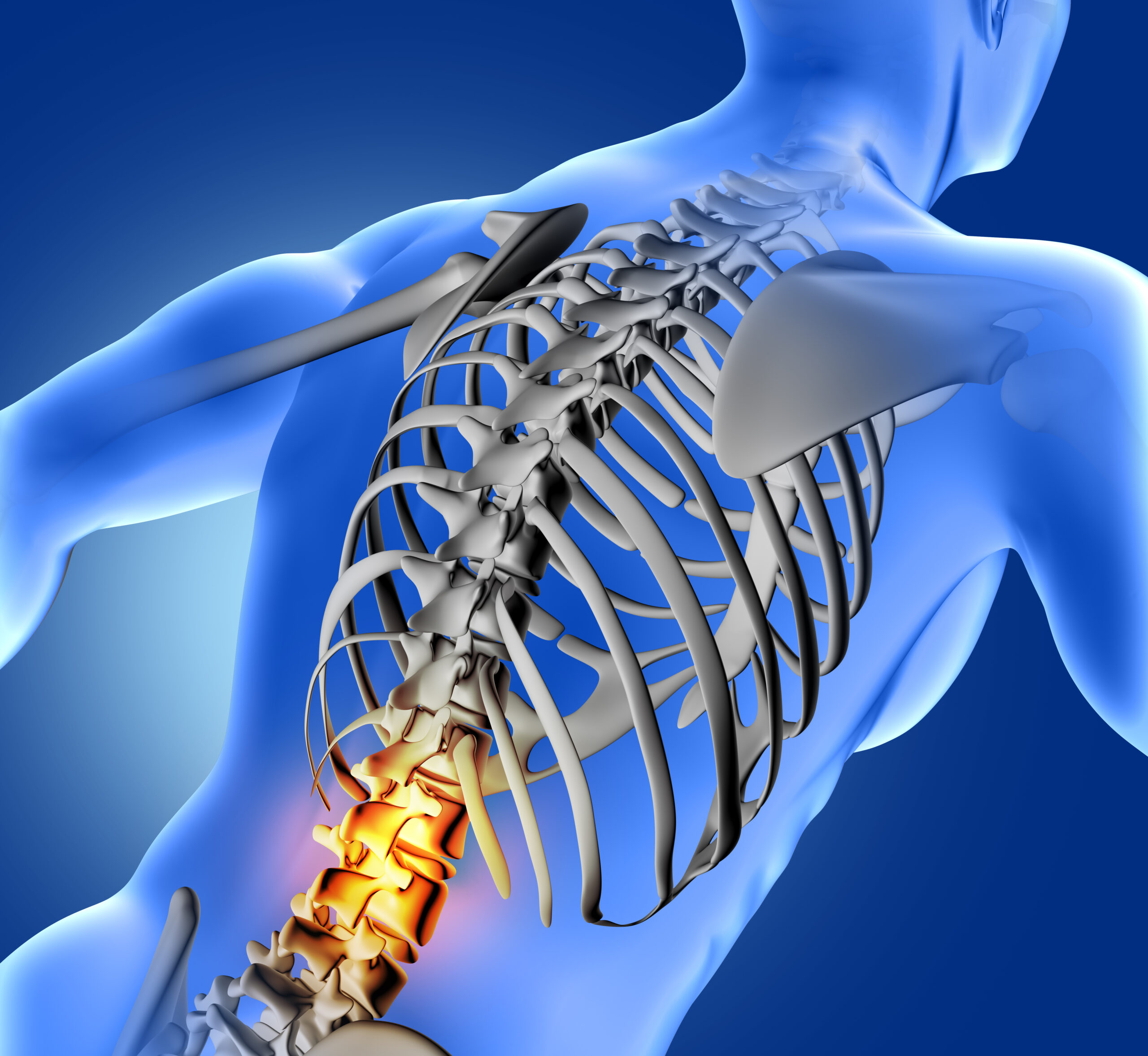
The spinal cord begins at the foramen magnum, where it continues seamlessly from the medulla oblongata of the brain. It travels downward through the protective vertebral canal and typically ends around the level of the first or second lumbar vertebra. On average, its length is about 45 cm in men and 43 cm in women.
Protected by tough layers called meninges and cushioned by cerebrospinal fluid, the spinal cord is suspended safely within the vertebral column. This design ensures both stability and flexibility while safeguarding one of the body’s most delicate structures.
Spinal Cord Segments
The spinal cord is divided into 31 segments, each giving rise to a pair of spinal nerves. These nerves emerge in an organized manner:
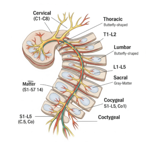
Cervical: 8 pairs
Thoracic: 12 pairs
Lumbar: 5 pairs
Sacral: 5 pairs
Coccygeal: 1 pair
Together, these nerves form an intricate network that connects the brain with every region of the body, enabling sensation and movement.
Inside the Spinal Cord
When viewed in cross-section, the spinal cord reveals two distinct areas: grey matter and white matter.
The grey matter sits at the center in a butterfly or H-shaped pattern. Its anterior horns house motor neurons responsible for movement, while the posterior horns contain sensory neurons that process incoming signals. The two sides of the grey matter are joined by the grey commissure, which surrounds a central canal filled with cerebrospinal fluid.
Encasing the grey matter is the white matter, made up of bundles of axons. These axons form columns (funiculi)—posterior, lateral, and anterior—that act as highways for communication between different parts of the spinal cord and the brain.
Nerve Branches
Each spinal nerve forms from the combination of two roots:
The posterior (dorsal) root, which carries sensory information from the body to the CNS.
The anterior (ventral) root, which sends motor commands from the brain to the body.
Once formed, spinal nerves split into branches. The dorsal ramus serves the muscles and skin of the back, while the ventral ramus extends to the limbs and the front of the body, transmitting both sensory and motor information.
Functions of the Spinal Cord
The spinal cord’s roles extend far beyond simply transmitting messages. Its main functions include:
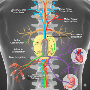
Signal transmission – carrying messages between the brain and body.
Reflex coordination – producing rapid, automatic responses to stimuli.
Basic integration – processing simple actions independently of the brain.
Autonomic regulation – helping manage vital processes like digestion and heart rate.
Communication pathways – serving as a hub for complex signaling between the CNS and the rest of the body.
Final Thoughts
The spinal cord is truly the body’s information highway, ensuring smooth communication and coordination. Without it, even the simplest movements or reflexes would be impossible. Understanding its structure and functions not only highlights the brilliance of the human body but also underscores why protecting spinal health is so essential.
want to be physical therapist-check Physioneeds Academy